|
|
|
1. Moderate mandibular atrophy. (Tutorial video presentation)
71- year old woman without retention of lower denture and moderate mandibular atrophy. Previously the denture was retained by the lower canines, which persisted as radices when patient was referred.
|
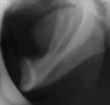
History of Parkinsons disease and receive medication for this. However the patient suffer severe tics in spit of the medication and a proper orthopantomographic radiograph was not possible to obtain. Preoperative radiographic evaluation was therefore restricted to a lateral radiograph.
Clinical and radiographic evaluation show that the residual height of mandibular body in the interforaminal region is 17-19 mm.
|
 |
Initially the radices of the canines were removed in local anesthesia and 6 weeks later the patient was taken to general anesthesia to have 4 implant inserted in the interforaminal region together with an immediate impression for a single stage, bar-supported overdenture.
|
|
01. Incision & Exposure mental foramen.
|
|
Incision on top of alveolar crest & releasing incision. The incision depth is superficial in the area of mental foramen until the exact position of the mental nerve is identified. The extent of the mucoperiostal flap is determined by the necessary osteotomy of alveolar crest and the need to identify the exact anatomy of residual ridge (i.e. lingual inclination).
|

|
|
|
|
|
|
|