|
|
|
|
|
Preoperative planning and examination, work-up of patient.
Seventy-two y. old lady with edentulous lower right mandibular segment distal to lower right canine. Last right second lower molar removed 3 month previously.
Upper left second molar removed two months previously.
In general good health record, no medications and no smoking habit. However patient has periodically experienced attacks of typical Trigiminal Neuralgia related to the left side of face (second and third branch).
- Examination:
- Smile line: Expose 2 mm of upper incisors at rest, 10 mm at smile & expose to first molar. Lower theeth hardly exposed.
- Occlusion stable, but are missing support in right side after loss of last second molar.
- Overjet horizontal 5 mm, vertical 4 mm. Inter-occlusal distance in edentulous area 5-6 mm.
Clinic slides:
Parodontal pockets up to 5 mm, Apical parodontal area first upper left bicuspid chronical infected with fistular lesion of oral mucosa. Oral mucosa in general without alterations.
Width of alveolar process at the surface clinical estimated to 5-6 mm.
Exact magnification of panoramic view is monitored by placing 5 mm balls in a wax impression.
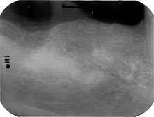
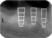
X-ray shows good height of residual alveolar process, i.e. 15 mm from top to mental foramen, 17 mm from top to inferior alveolar nerve region first lower molar. The distance between anterior ‘knee’ of alveolar nerve and the surface of right lower cuspid is 6 -7 mm.
|
|
|
 |
 |
|
|
|
|
|